
At proteins are macromolecules formed by the successive union of amino acids, which are compounds originated from the peptide bond among a group amino and a group carboxylic. This definition is well explained in the text. Chemical Composition of Proteins.
Primary structure of proteins
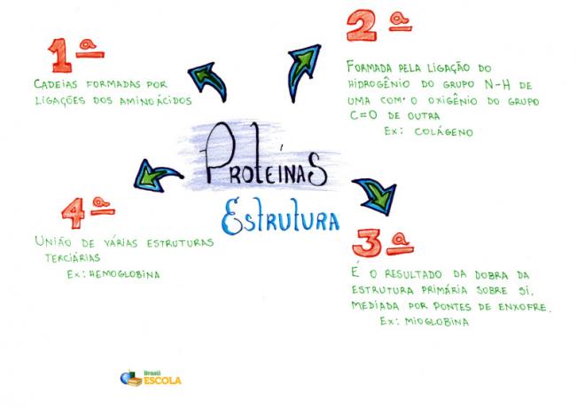
The main chain of the protein formed by the binding of amino acids and that shows the sequence in which they appear is called the primary structure of the protein.

Model of the primary structure of a protein
However, the same protein can also acquire secondary, tertiary and even quaternary structures. This occurs as a result of intermolecular interactions between parts of the same protein or between several protein chains.
Mind Map: Proteins
* To download the mind map in PDF, Click here!
Protein secondary structure
The secondary structure is usually the result of hydrogen bonds that occur between the hydrogen of the –NH group and the oxygen of the C═O group. Thus, structures like the ones shown below are formed, which are similar to a spring (an example occurs with the keratin from our hair) or as folded sheets of paper (this type occurs with the fibroin of the web of the Spider):

Examples of protein secondary structures
These are just two examples of possible secondary structures of proteins. Below is the secondary structure of collagen. The interactions that resulted in a spiral-shaped structure are hydrogen bonds:

Collagen secondary structure
Tertiary structure of proteins
When the primary structures of proteins fold back on themselves, they give rise to a spatial arrangement called a tertiary structure. It usually occurs as a result of sulfur bonds known as disulfide bridges. But other spatial bonds can also occur, such as those made by metal atoms.
The following is the tertiary structure of myoglobin:

Tertiary structure of myoglobin
Quaternary structure of proteins
The quaternary structure, on the other hand, is the union of several tertiary structures that assume well-defined spatial forms. Here's a model of the quaternary structure of human hemoglobin, the protein in red blood cells that carries oxygen around the body:

Quaternary structure of hemoglobin
This structure is formed by four tertiary structures. Among them are prosthetic groups (heme) formed by iron, as shown in the following illustration:

Structural formula of Heme B, component of hemoglobin
* Mind Map by Me. Diogo Lopes Dias
By Jennifer Fogaça
Graduated in Chemistry
Source: Brazil School - https://brasilescola.uol.com.br/quimica/estruturas-das-proteinas.htm