
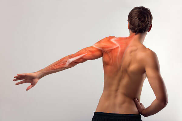
O muscular system is the system formed by muscle set of our body. They correspond to about 50% of the total weight of our body, and the contraction of these structures is responsible for several functions, among which we can highlight the movement.
Read too:Amyotrophic lateral sclerosis — a disease that causes muscles to atrophy
Muscle Functions
Our body is a complex machine that features several integrated systems working together to ensure our survival. Among these systems, the muscular one stands out, which is related to important functions of the body. Check out some of your main functions:
They guarantee the movement of the body.
They promote the stabilization of body positions.
They are responsible for moving blood through the body, food through the digestive system and of the urine fur urinary system.
They guarantee the realization of the breathing movements.
Read too: What are peristaltic movements?
types of muscles
The human body is formed by
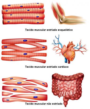
threemuscle types different: the skeletal striatum, the cardiac striatum and the non-striatum. You musclesstriatedskeletal are normally associated with the systemskeletal and have only movementvoluntary, that is, its contraction is conscious. The term striatum is associated with the fact that these muscles have light and dark bands, which are arranged alternately when observed under optical microscopy.You striated cardiac muscles, as the name implies, are exclusive of the heart. They have a striated appearance, like the skeletal one, but they have contractionsinvoluntaryand vigorous.
You unstriated muscles, in turn, present involuntary and slow contraction and are found in the digestive and respiratory systems, as well as in some hollow structures such as the urinary bladder and small intestine. One of its most striking features is the absence of striations, which is observed in other muscle types.
Characteristics of muscle tissue | |||
Feature |
Skeletal striated muscle tissue |
Cardiac striated muscle tissue |
Unstriated muscle tissue |
cells |
Long cylindrical cells, multinucleated and with striations |
Elongated and branched cells, with only one or two nuclei and striations |
Spindle cells, with a single nucleus and without striations |
Contraction |
volunteer |
involuntary |
involuntary |
Location |
bones |
Heart |
Digestive system, bladder, arteries and other internal organs |
skeletal muscle contraction
the human body has more than 600 skeletal muscles, that present contractionvoluntary. These muscles are formed by elongated, multinucleated cells, which are also called muscle fibers. One of the striking characteristics of this type of muscle tissue is the presence of transverse striations.
At muscle fibers have filaments of myosin and actin, that are proteins with the ability to contract. Actin and some other proteins that are associated form so-called fine filaments. Myosin forms the thick filaments. The thin and thick filaments alternate, forming light and dark bands.
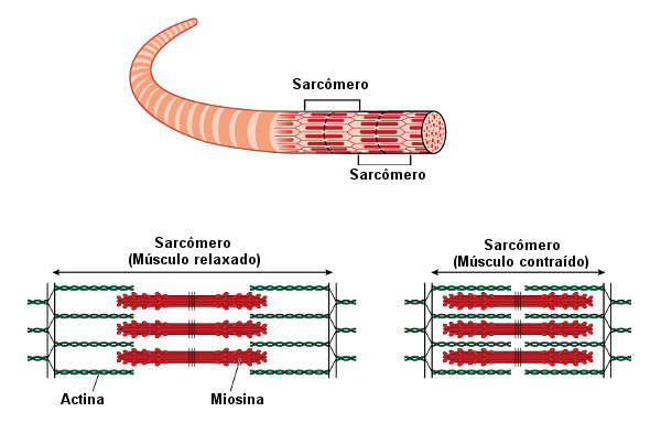
The light bands are formed by thin filaments and are called I bands. They are so called because they are isotropic under a polarizing microscope. The dark bands are called bands A, as they are anisotropic under a polarizing microscope and are characterized by the presence of thin and thick filaments.
In the center of band I there is a dark line, called Z line. It delimits the sarcomere, which is formed by two halves of I bands and a central A band. In the center of the A band, we have the H band, a lighter region where only myosin filaments are found.
In muscle contraction occurs the shortening of the sarcomeres and, consequently, of the entire fiber. During contraction, actin and myosin filaments overlap, which makes the I and H bands narrower. To learn more about this process, be sure to read our text: Contraction in skeletal muscles.
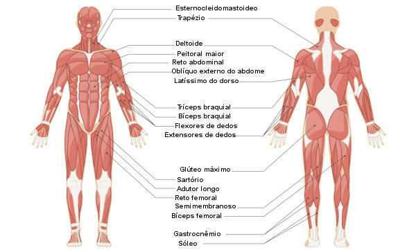
Major skeletal muscles
There are hundreds of skeletal muscles in our bodies, each performing a certain function. These muscles can be placed in large groups, these are:
Muscles of the face and scalp: examples: orbicularis oculi and levator labii superioris.
Muscles of mastication: examples: masseter and medial pterygoid.
Abdominal wall muscles: examples: internal oblique and transverse abdomen.
Muscles that move the head and shoulder: examples: trapeze and elevator of the scapula.
Muscles that move the spine: examples: long chest and long neck.
Muscles that move the tongue: examples: genioglossus and hyoglossus.
Muscles that move the hip and knee joints: examples: gluteus maximus and abductor longus.
Muscles that move the forearm: examples: triceps and biceps.
Muscles that move the foot and toes: examples: flexor digitorum longus and abductor hallucis.
Muscles that move the thumb: examples: extensor pollicis longus and extensor pollicis brevis.
Muscles that move the wrist: examples: flexor carpi radialis and extensor carpi radialis brevis.
Muscles that move the humerus: examples: deltoid and supraspinatus.
Muscles that move the fingers: examples: flexor digitorum profundus and extensor index finger.
Respiratory Muscles: examples: diaphragm and external intercostals.
Supra and infrahyoid muscles of the neck: examples: myloioid and genioioid.
Read too:What is the diaphragm?
→ Classification of skeletal muscles
When we study the muscular system, we see the classification of the skeletal muscles that make up our body. This is due to the fact that the non-striated muscles are part of the organs and normally do not receive their own name, as well as the striated cardiac muscle, which is present in the heart.
There are over 600 skeletal muscles in our body, which represents about 50% of our entire body mass. They are classified based on various criteria, such as their origin and insertion, action, function, fiber shape and arrangement, and number of heads.
it is understood by origin the place where the muscle is most attached and which serves as the basis for its action. already the insertion it is the moving point at which it is possible to observe the effect of movement. The gluteus minimus, for example, is a muscle responsible for the abduction of the thigh and originates from the lateral surface of the ileum. Its insertion is on the anterior surface of the femur, more precisely in the region of the greater trochanter (a prominence located on the upper edge of the femur).
When muscles are classified according to their action, they are called extensors, flexors, adductors, abductors, rotators, supinators and pronators. See the function of each one:
Extenders: stretch a limb;
Flexors: are responsible for bending;
Adductors: lead a limb towards the midline of the body;
Abductors: move the member off that line;
Rotators: rotate the limbs;
Supinators: turn the palm up;
Pronators: put the palm down.
Observing the function, the muscles can be classified into agonists, antagonists and synergists. Agonist muscles are directly responsible for the desired movement, being the main agents in the execution of a movement; antagonists are muscles that offer resistance to the action of the agonist muscle; and synergists are muscles that assist antagonists, ensuring that excessive movement does not occur.
Regarding the shape and arrangement of fibers, muscles can be classified into parallel fiber or oblique fiber muscles to the direction of traction exerted by them. As an example of parallel fiber muscles, we can mention the biceps and pectoral muscles. As an example of an oblique fiber muscle, we can mention the extensor longus of the toes.
Finally, when the criterion used is number of heads, it takes into account how many tendons of origin the muscle has. The biceps, for example, has two heads; the triceps, three; and the quadriceps, four.
By Ma. Vanessa dos Santos
Source: Brazil School - https://brasilescola.uol.com.br/biologia/sistema-muscular.htm