Muscle tissue it is a type of animal tissue that presents as its most striking feature its ability to contract. This tissue is essential for the functioning of our body, being responsible for ensuring, for example, our movements and the beat of the heart.
Read too:Muscle system - definition, function and types of muscles
Muscle Tissue Characteristics
Muscle tissue presents cells with capacity of contraction, being also called muscle fibers. These cells, or fibers, are elongated and have a large amount of contractile protein filaments, such as actin and myosin.
Some muscle cell structures have specific names. THE membrane cell, for example, is called sarcolemma. O cytosol presents the denomination of sarcoplasm. already the endoplasmic reticulum smooth is styled sarcoplasmic reticulum.
Importance of muscle tissue
Due to its ability to contract, extensibility, elasticity and excitability, muscle tissue plays an important role in our body. It is due to his presence that we are able to:
Move our body;
Ensure the heartbeat;
Allow the movement of various substances, such as blood and the food;
Ensure the stabilization of the body and maintenance of posture;
Allow some organs to grow in size and return to their original size;
Produce heat by its contraction.
Do not stop now... There's more after the advertising ;)
Classification of muscle tissue
Muscle tissue can be divided into three groups: skeletal striated muscle tissue, non-striated muscle tissue, and cardiac striated muscle tissue. We'll talk a little more about each of these types:
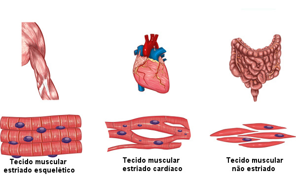
Skeletal striated muscle tissue
Features long, cylindrical and multinucleated cells, which are located on the periphery of them. As the name suggests, this tissue has striations, which are transverse. Its cell bundles are long and can reach up to 30 cm, while the fiber diameter varies between 10 µm to 100 µm.
Each muscle fiber has a series of bundles of filaments, the myofibrils. Four proteins different ones are found in these myofibrils (myosin, actin, tropomyosin and troponin), and myosin and actin are the most abundant.
See too:Chemical composition of proteins
When viewed under an optical microscope, skeletal muscle fibers show alternating light and dark bands which guarantees the pattern of transverse striations. The dark band is called band A and it is formed by thin (actin) and thick (myosin) filaments, while the light band is called band I and is formed only by thin filaments.
In the center of each band I, there is the presence of a dark transversal line, called Z line, which delimits the so-called sarcomere. Band A has a lighter region in the center, called band H, which is formed only by thick filaments.

In these muscles there is a repetition of units called sarcomeres, which are repetitive and basic units of the myofibrils of this type of muscle. Each sarcomere is made up of the part that lies between two successive Z lines: two I-band halves and an A-band in the center.
THE contraction of this type of tissue is rapid and vigorous, but not involuntary. For the contraction of these cells to occur, our commands are fundamental, and in this process there is a shortening of the sarcomeres. The striated skeletal muscles are attached to our bones, ensuring that muscle contraction is converted into movement. As an example we have the biceps it's the triceps.
Read too: Human skeleton - name of bones, functions and divisions
Cardiac striated muscle tissue
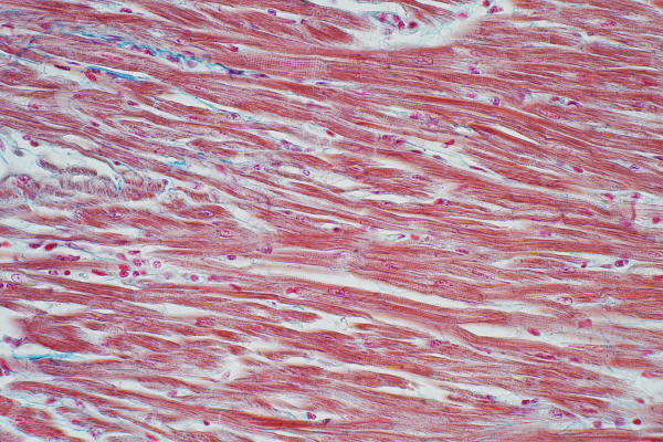
It is located in the heart and, like the previous one, it has striations in its cells. They are elongated and branched, these branches being joined by structures called intercalary disks.
These discs transmit signals from one cell to another, guarantee the synchronization of the cardiac contraction and act by preventing the cells from separating when the heart beats. Unlike skeletal muscle tissue, cardiac muscle fibers have one or two cores, which are in the most central position or close to that region.
The striated cardiac muscle contractions are involuntary, that is, they happen independently of our control. In this type of muscle, the cells have a series of mitochondria,which is easy to understand, since they have high metabolism and constantly need ATP.
Know more: Cardiovascular system - anatomy, function, organs and summary
Muscle tissue not striated or smooth
It is formed by cells that do not have striations, this being a feature that allows easy differentiation from other types of tissue. Its cells are long, with a thicker center and tapered ends. They have only a single nucleus, arranged in the center of each one of them.
Unstriated muscle tissue presents involuntary contraction, not vigorous as seen in other tissues. The contraction is controlled by the nervous system autonomous. This tissue is found forming the walls of various internal organs, such as those of the digestive tract, the bladder and even the arteries.

By Vanessa Sardinha dos Santos
Biology teacher

