O human heart, as well as all mammals, it is a organmuscular formed by four cavities and whose primary function ensure that the blood be sent for all parts of our body. This organ, which is about the average size of a clenched fist, is located behind the sternum and between the lungs, in the middle mediastinum. It guarantees pumping thanks to the rhythmic contractions of the heart muscle.
Read too:Know the entire cardiovascular system
heart anatomy
The heart, an organ of the cardiovascular system, is a hollow chamber with four cavities: two atria and two ventricles. It is shaped like an inverted cone with the apex facing downwards and has the approximate volume of a clenched hand. It usually weighs around 300 g.
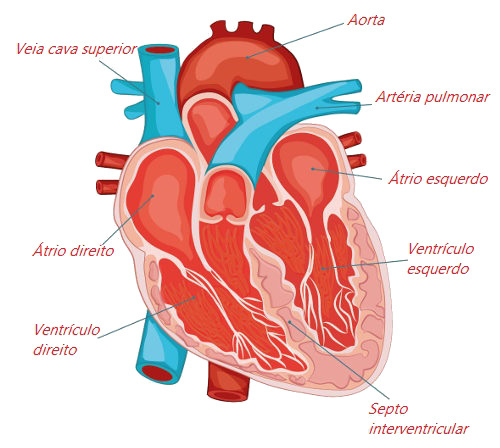
Look at the main parts of the human heart.
The walls of the heart are composed of three tunics:
endocardium: tunica formed by endothelium that rests on a layer of loose connective tissue, the subendothelial layer. This last layer is connected to the myocardium by the subendocardial layer, which has nerves, veins and branches of the system that conducts the nerve impulse.
Myocardium: thickest layer of the heart and responsible for the organ's ability to contract. This layer is formed by bundles of cardiac muscle fibers, which are oriented in several directions. The myocardium surrounds the chambers of the heart, being thicker in the ventricles – especially in the left ventricle – and thinner in the walls of the atria.
Pericardium: invaginated sac surrounding the heart and formed by the parietal pericardium and the visceral pericardium. The parietal pericardium is an outer layer, while the visceral pericardium is an inner layer. The visceral pericardium forms what is called the epicardium and lines the heart externally.
heart chambers
The heart is formed by two upper chambers, the atriums, and two lower chambers, the ventricles. The atria are separated by the interatrial septum, and the ventricles are separated by the interventricular septum.
The atriums have as their main function receive the blood which comes from different parts of the body, thus functioning as collecting chambers. The ventricles are responsible for ensuring the pumping blood to other locations, being, therefore, pump chambers.
Given the role of each heart chamber, it is easy to understand why the ventricles have more developed walls, and the atria, thinner walls. At wallsthick provide a contraction much more vigorously than the thin walls, thus ensuring that blood is sent to different parts of the body.
The right atrium is a chamber that receives blood from different parts of the body except the lungs. Three veins flow into it: superior vena cava, inferior vena cava and coronary sinus. The right ventricle communicates with the right atrium and part of the right atrium. pulmonary artery, which takes blood to the lungs.
The left atrium receives blood from the lungs through four pulmonary veins. The left ventricle receives blood from the left atrium and leaves the aorta artery, which is responsible for carrying blood to the rest of the body, except the lungs.
Read too: Vertebrate heart – see the differences between groups
heart valves

Note the four valves found in the heart.
Between the atria and the ventricles, we find the atrioventricular valves. In the heart, the semi-lunar valves pulmonary and aortic, located between the ventricles and the pulmonary artery and aorta, respectively. Therefore, we have four valves in the heart, which prevent the backflow of blood, thus providing a movement of blood in a single direction.
O heart murmur it is nothing more than an abnormal sound heard between beats that is the result of turbulence in the blood flow. The heart murmur does not always determine a pathology, being called innocent heart murmur the one that occurs even without anatomical and/or functional alterations of the cardiovascular system. In other situations, the murmur may be the result of some problems, such as the passage of blood through an abnormal valve structure. |
the way of blood in the heart
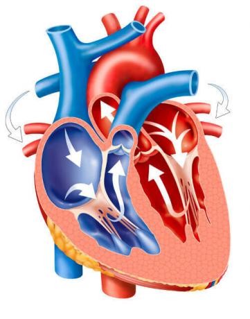
Look closely at the arrows and notice the path taken by the blood inside the heart.
O bloodhe arrives of the different parts of the body, except the lung, by the right atrium. Into this cavity, the veinsarmholes upper and lower, which bring blood from the body, and also the coronary sinus, which is responsible for draining the blood present in the heart itself.
From the atrium the blood flows to the right ventricle and is carried to the lungs via the pulmonary artery. After suffering hematosis in the lungs, the blood is carried through four pulmonary veins to the left atrium, from where it flows to the Left ventricle. From this last cavity, blood is driven out through the aorta artery, which carries blood to all parts of the body, except the lung, through numerous branches.
Read too:Blood vessels - what they are, types, function, structure
heart impulse
The heart beats rhythmically, alternating between contraction and relaxation. THE contraction is called systole, it's the relaxation is called diastole. Systole ensures that blood is pumped, and diastole, which is the relaxation phase, ensures that the heart's cavities fill with blood.
The generation of the heartbeat is attributed to the call sinoatrial node, which is formed by a mass of specialized cells, located in the wall of the right atrium, responsible for spontaneously generating a electric impulse. These electrical impulses are similar to generated by the nerve cells and propagate quickly through the muscle tissue cardiac.
These impulses reach another point that guarantees the transmission of impulses: the atrioventricular node, which is located between the wall of the two atriums. The signals then depart from the atrioventricular node and travel towards all parts of the ventricles and the apex of the heart through specialized structures called the Purkinje system.
Did you know that the average normal heart rate is approximately 70 beats per minute. When this rhythm is too high, there is tachycardia, and when it is too low, brachycardia. |
Read too:10 leading causes of death in the world
World Heart Day
O World Heart Day is celebrated on the day September 29th and aims to alert the population of the need to take care of this important organ of our Cardiovascular system. The creation of the date, according to the Ministry of Health, was motivated by the fact that cardiovascular diseases are the main cause of death in the world. Therefore, World Heart Day is an important moment for reflect about our life habits and how can we improve the health of our heart.
By Ma. Vanessa dos Santos
Source: Brazil School - https://brasilescola.uol.com.br/biologia/coracao-humano.htm