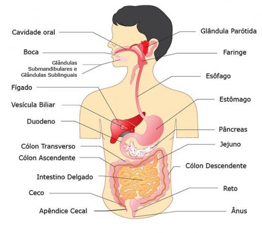
O Digestive system is also known as Digestive System or digestive system. It is formed by a set of organs that act in the human body.
The action of these organs is related to the food transformation process, which aims to help in the absorption of nutrients.
All of this happens through mechanical and chemical processes.

Digestive System Components
The Digestive System (new nomenclature) is divided into two parts.
One of them is the digestive tube (properly said), formerly known as the digestive tube. It is divided into three parts: high, medium and low. The other part corresponds to the adjoining organs.
See in the table below the organs that make up each part of the Digestive System.
| parts | Description |
|---|---|
| upper digestive tube | Mouth, pharynx and esophagus. |
| Middle digestive tube | Stomach and small intestine (duodenum, jejunum and ileum). |
| Low digestive tube | Large intestine (cecum, ascending colon, transverse, descending, sigmoid curve and rectum). |
| Attached bodies | Salivary glands, teeth, tongue, pancreas, liver and gallbladder. |
More information and details about each of the components of the Digestive System are presented below.
Upper Digestive Tube

The upper digestive tube is formed by the mouth, pharynx and esophagus.
Find out more details about each of these organs below.
Mouth

The mouth is the gateway for food into the digestive tract. It corresponds to a cavity lined with mucosa, where food is moistened by the Spittle, produced by the salivary glands.
In the mouth, chewing occurs, which corresponds to the first moment of the mechanical digestion process. It happens with teeth and tongue.
In a second moment, the enzymatic activity of ptyalin, which is salivary amylase, comes into play. She acts on the starch found in potatoes, wheat flour, rice and transforming it into smaller molecules of maltose.
Pharynx

THE pharynx it is a membranous muscular tube that communicates with the mouth, through the isthmus of the throat and at the other end with the esophagus.
To reach the esophagus, the food, after being chewed, travels through the entire pharynx, which is a common channel for the digestive system and the respiratory system.
In the swallowing process, the soft palate is retracted upwards and the tongue pushes food into the pharynx, which voluntarily contracts and carries the food into the esophagus.
The penetration of food into the airways is impeded by the action of the epiglottis, which closes the communication orifice with the larynx.
Esophagus

O esophagus it is a muscular conduit, controlled by the autonomic nervous system.
It is through waves of contractions, known as peristalsis or peristaltic movements, the muscular duct squeezes food and takes it towards the stomach.
You may also be interested in:
- Muscle tissue
- Nervous system
Medium Digestive Tube
The middle digestive tube is formed by the stomach and small intestine (duodenum, jejunum and ileum).
Learn about each of them below.
Stomach

O stomach it is a large pocket that is located in the abdomen, being responsible for the digestion of proteins.
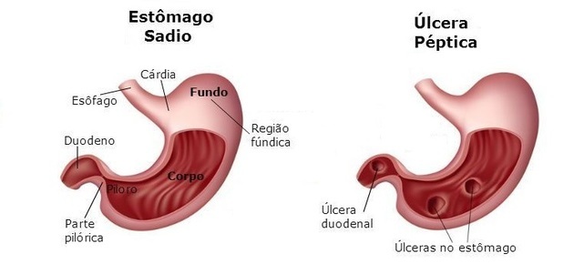
The entrance to the organ is called the cardia, because it is very close to the heart, separated from it only by the diaphragm.
It has a small upper curvature and a large lower curvature. The more dilated part is called the "fundic region", while the final part, a narrow region, is called the "pylorus".
The simple movement of chewing food already activates the production of hydrochloric acid in the stomach. However, it is only with the presence of food, which is protein in nature, that the production of gastric juice begins. This juice is an aqueous solution, made up of water, salts, enzymes and hydrochloric acid.
The gastric mucosa is covered by a layer of mucus that protects it from the aggression of the gastric juice, since it is very corrosive. Therefore, when an imbalance in protection occurs, the result is an inflammation of the mucosa (gastritis) or the appearance of wounds (gastric ulcer).
THE pepsin it is the most potent enzyme in gastric juice and is regulated by the action of a hormone, gastrin.
Gastrin is produced in the stomach itself when protein molecules from food come into contact with the wall of the organ. Thus, pepsin breaks down large protein molecules and turns them into smaller molecules. These are proteoses and peptones.
Finally, the digestion gastric pain lasts, on average, two to four hours. In this process, the stomach undergoes contractions that force the food against the pylorus, which opens and closes, allowing the chyme (white foamy mass) to reach the intestine in small portions slender.
Small intestine

O small intestine it is covered by a wrinkled mucosa that has numerous projections. It is located between the stomach and the large intestine and has the function of secreting the various digestive enzymes. This gives rise to small, soluble molecules: glucose, amino acids, glycerol, etc.
The small intestine is divided into three parts: the duodenum, the jejunum and the ileum.
O duodenum it is the first portion of the small intestine to receive the chyme that comes from the stomach, which is still very acidic, being irritating to the duodenal mucosa.
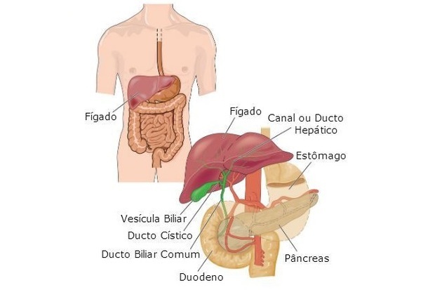
Soon after, the chyme is bathed in the bile. Bile is secreted by the liver and stored in the gallbladder, containing sodium bicarbonate and bile salts, which emulsify lipids, fragmenting their drops into thousands of micro droplets.
In addition, the chyme also receives pancreatic juice, produced in the pancreas. It contains enzymes, water and large amounts of sodium bicarbonate, as this favors neutralization of the chyme.
Thus, in a short time, the duodenal food “pape” becomes alkaline and generates the necessary conditions for intra-intestinal digestion to occur.
already the jejunum it's the ileum they are considered the part of the small intestine where the bolus transit is fast, remaining empty most of the time during the digestive process.
Finally, along the small intestine, after all the nutrients have been absorbed, there is a thick paste made up of unassimilated debris and bacteria. This paste, already fermented, goes to the large intestine.
Low Digestive Tube
The lower digestive tube is formed by the large intestine, which has the following components: cecum, ascending colon, transverse, descending, sigmoid curve, and rectum.
Large intestine

O large intestine it measures about 1.5 m in length and 6 cm in diameter. It is a place for water absorption (both ingested and digestive secretions), storage and elimination of digestive waste.
It is divided into three parts: the cecum, colon (which is subdivided into ascending, transverse, descending, and the sigmoid curve) and rectum.
In the cecum, the first portion of the large intestine, food waste, already constituting the “fecal bun”, passes to the ascending colon, then to the transverse and then to the descending colon. In this portion, the fecal bolus remains stagnant for many hours, filling the portions of the sigmoid curve and rectum.
The rectum is the final part of the large intestine, which ends with the anal canal and anus, through which feces are eliminated.
To facilitate the passage of the fecal bolus, the glands in the mucosa of the large intestine secrete mucus in order to lubricate the fecal bolus, facilitating its transit and elimination.
Note that vegetable fibers are not digested or absorbed by the digestive system, they pass through the entire digestive tract and form a significant percentage of the fecal mass. It is, therefore, important to include fiber in the diet to help with the formation of feces.
You may also be interested in:
- Appendix
- human body systems
- Organs of the human body without which you can survive


