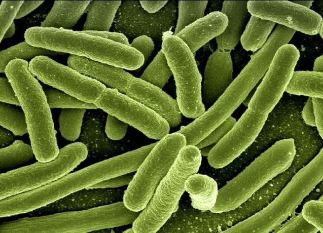
O trypanosoma cruzi is a single-celled flagellate protozoan that causes Chagas disease.
O T. crossed it is characterized by the presence of a single flagellum, a large mitochondria, and the kinetoplast, a compartment in the mitochondrion that contains DNA.
The geographic distribution of T. crossed it extends from the southern United States to southern Argentina. In this region, there are several cases of Chagas disease.
Chagas disease is transmitted through the feces of an insect, the barber, which contains the infective forms of the T. crossed.
Learn more about Chagas disease.
Morphology
During its life cycle, the T. crossed it can present three morphological forms: amastigote, epimastigote and trypomastigote.
- amastigot: presents a rounded shape. The nucleus and kinetoplast are not seen with light microscopes. It has no scourges. Present in the intracellular phase, during the chronic phase of the disease.
- epimastigote: Variable size with elongated shape and semi-central core. It represents the form found in the barber's digestive tract, the vector of Chagas disease.
- Trypomastigote: Has an elongated and spindle-shaped “c” or “s” shape. It is the form present in the extracellular phase, which circulates in the blood, in the acute phase of the disease. It is the infective form for vertebrates.
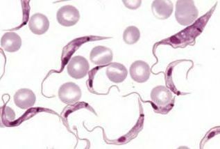
Trypanosoma cruzi in its trypomastigote form in the blood
Life cycle
the life cycle of T. crossed it starts when the barber, when feeding on the vertebrate host, eliminates its feces and urine, where trypomastigote forms may be present.
Trypomastigote parasites penetrate the skin and infect host cells, where they transform into the amastigote form.
When cells are full of parasites, they again change to the trypomastigote form. Because they have a large amount of parasites, the cells break up and the protozoa reach the bloodstream, reaching other organs.
At this stage, if the vertebrate host is bitten by the barber, the protozoa will be transmitted to the insect. In the barber's intestine, they change their shape to epimastigotes, where they multiply and become trypomastigotes again, the forms infecting vertebrates.
Know more:
Protozoa
Diseases caused by protozoa