THE leaf it is a organvegetable, usually laminar and green, found in majorityof theplants existing. It usually has limited growth, that is, it does not grow throughout the life of the plant. Its main function is the photosynthesis, but also works in perspiration, gas exchange, reserve and even in the attraction ofpollinator agentss. Next, we will learn more about the anatomy of this important plant organ.
Read too:Photosynthesis: process carried out by producers in the food chain
plant leaf anatomy
The sheet presents twofaces, The adaxial face (top) and the abaxial face (lower part). Both sides are covered by the epidermis, which is characterized by being continuous throughout the sheet. In addition to this fabric, we can clearly see the presence of fabricsvascular running through the entire sheet, forming the calls ribs. These branch off fur mesophyll, which constitutes the fundamental system of the leaf. Therefore, we realize that this organ is formed by a dermal system (epidermis), a fundamental system (mesophyll) and a vascular system (vascular bundles).

Epidermis
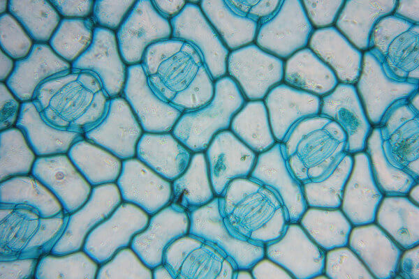
THE epidermis is the fabric that covers the entire sheet, being generally formed by only one layer, with the exception of some species, such as those of the genus Piper. In the epidermis, we observe the presence of cells performing different functions, but most of the tissue is formed by ordinary epidermal cells, which are tightly arranged.
Among common epidermal cells, we can observe the presence of other specialized cells, for example, the cells that form the stomata and trichomes — two structures that can occur on both sides of the sheet or on just one of them.
Generally, the term is used stomato to refer to the complexstomatal, formed by two guard cells that delimit the stomatal cleft (ostiolus). The main function assigned to this structure is the performing gas exchange.
You trichomes they are also important structures found in leaves. These epidermal appendages have different shapes and can be classified into ceilings or in glandular. We can differentiate these trichomes quite easily, since only the trichomesglandular are related to substance secretion.

In the cell wall of epidermal cells, we also observe the presence of cutina, a lipid compound. The cutin can be impregnated to the wall or form a layer called cuticle on top of the epidermis.
The cuticle is mainly related to protection against excessive water loss. The presence of wax is also verified, which may be deposited on the cuticle or within the cutin matrix.
Read too:Kingdom Plantae in Enem
mesophyll
O mesophyll, leaf tissue responsible mainly for the photosynthesis process, is basically formed by parenchyma chlorophyllan, but we can also find collenchyme and sclerenchyma. O parenchymachlorophyll characterized by having a large amount of chloroplasts. When making a transversal cut on the leaf blade, we can see it between the epidermis of the abaxial and adaxial faces.
In the eudicots we can often observe the presence of two main types of chlorophyllian parenchyma: palisade and spongy.
O palisade parenchyma it may be located below the epidermis of the adaxial face or on both sides. It is characterized by having elongated cylindrical cells, arranged side by side and separated by small intercellular spaces.
already the spongy parenchyma has a large amount of space between the cells, and these have a varied format. It is located just above the epidermis of the abaxial face or between the layers of palisade parenchyma.

A sheet is called dorsiventral when the palisade parenchyma is close to the adaxial epidermis, and the spongy parenchyma is close to the abaxial surface. When the palisade parenchyma is located on both the abaxial and adaxial sides, we say that the leaf is isolateral.
There are also leaves called homogeneous, in which it is not possible to differentiate these two types of parenchyma. In corn and some other grasses, it is not possible to check this differentiation.
vascular system
The leaf mesophyll is penetrated by several bundlesvascular, which are formed by xylem and by phloem. The vascular system of the leaves is continuous with the vascular system of the stem and, as said, can be called the ribs.
In the vast majority of leaves, we can observe the bundles with the xylem facing the adaxial face and the phloem facing the abaxial face. When the bundles are presented in this way, they are called collateral. The vascular bundles are surrounded by parenchymal cells, which form the so-called beam sheath. This sheath prevents vascular tissues from being exposed to the intercellular spaces present in the mesophyll.
The ribs are arranged differently on each sheet type. In eudicots, for example, the pattern of reticulated venation, in which ribs of smaller caliber appear based on a larger one. In most monocots, the ribs are parallels and have similar gauges.
Read more:Biology concepts that should not be confused in Enem
Importance of studying leaf anatomy
The anatomical features of a leaf can tell us a lot about a plant. Based on anatomical studies, we can, for example, obtain data about how the different luminosities and seasons of the year can cause changes in these organs and how these variations help the survival of the plant. In addition, leaf anatomy can provide important data for the taxonomy of groups, helping to identify microscopic differences between species.
By Ma. Vanessa dos Santos
Source: Brazil School - https://brasilescola.uol.com.br/biologia/anatomia-folha-vegetal.htm

