At veins, as well as the arteries, they are blood vesselss. For a long time, these vessels were defined as blood vessels that carried blood rich in carbon dioxide, also called venous blood. However, this is not true, as the pulmonary veins are responsible for carrying oxygen-rich blood, called arterial blood. The best definition of a vein, therefore, is one that presents it as a blood vessel whose function is to ensure that the blood, present in several fabrics of the body, return to the heart.
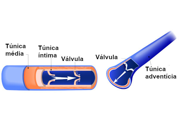
the veins have a wall formed by three layers: tunica intima, tunica media and tunica adventitia. This wall is less developed than the arterial wall, being about 1/3 the thickness of an artery wall. The veins carry blood at low pressure, and, to guarantee the return of blood to the heart, they have valves. In certain situations, these valves can present problems, triggering the emergence of varicose veins.
Read too: Difference between vein, artery and capillary
Vein characteristics
Veins are important blood vessels that bring blood back to the heart. They can be classified, according to their caliber, in: small, medium and large, being the
most of them small or medium caliber, with a diameter of 1 mm to 9 mm. Veins have three layers forming their walls, as well as arteries. The wall of veins, however, differs from that of arteries in that it is thinner. In arteries, the presence of thick walls is essential to support high blood pressure, which is not observed in veins, in which blood flows at low pressure. The layers that form the wall of the veins are:
Underwear: is an inner layer and is formed by cells endothelial cells, which are supported by a connective tissue loose. This layer is usually thin. However, in large-caliber veins, the tunica intima is well developed.
Middle tunic: in it we observe, mainly, cells of the muscle tissue smooth. A matrix with different components, such as elastic and reticular fibers, is also observed. It is noteworthy that, when compared to the tunica media of the arteries, this layer has fewer muscle cells and also a smaller amount of elastic fibers.
Adventitious tunic: in it the presence of mainly elastic fibers and collagen. It is a more developed layer in veins than in arteries.
As mentioned, veins are blood vessels that have the function of returning blood from tissues to the heart. This return is not always easy, since, in these vessels, the blood is under low pressure and, many times, the return must happen against the action of gravity.
To ensure this return, the veins have valves that help to maintain the unidirectional flow, thus avoiding the blood reflux. These valves are folds of the tunica intima that project into the vessel and have a crescent shape. In addition to the action of the valves, the vein guarantees the return of blood thanks to the smooth muscle contractions present in its wall and the contractions of the skeletal muscles that surround the vein.
Venules

Venules are vessels that collect blood from the capillaries and have a diameter of 0.1mm to 0.5mm. They form through the fusion of capillaries and progressively join together to form veins.
Read more: Cardiovascular system - responsible for the circulation of blood in our body
classification of veins
Veins can be classified as deep and superficial. At superficial veins are those that transit above the muscular fascia and can be seen through the skin. They are more caliber in the limbs and neck. Due to their location, they can be used as access routes for punctures. In people with developed musculature, these veins can be seen easily.
At deep veins, in turn, they are located more internally, being found transiting below the muscular fascia. They can be arranged to follow the arteries or stand alone.
Read more: Human heart - ensures that blood reaches all parts of our body
Varicose veins and blood circulation in the veins
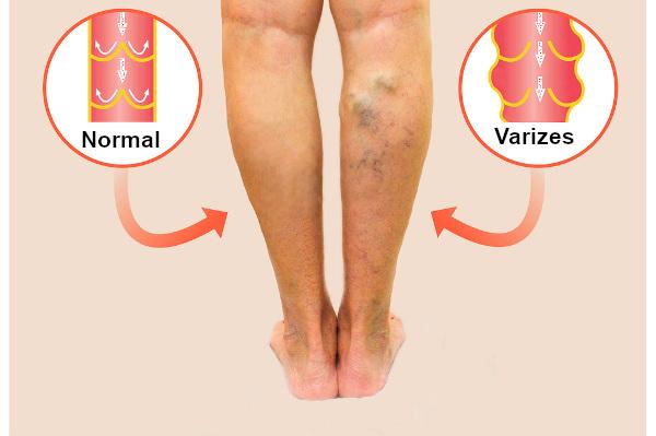
Veins are blood vessels through which blood circulates under low pressure, making unidirectional flow difficult. The valves in the veins act, in this context, preventing blood reflux. In certain situations, however, these valves are defective, which ends up harming the blood flow and causing dilations in the veins due to accumulated blood. These veins, called varicose veins, appear tangled and tortuous.

Varicose veins commonly appear in the legs, and the development of varicose veins is related, for example, to the standing for long periods.
teachers they are strong candidates for developing the problem due to the fact that, during work, they move little, remaining for long periods standing or sitting. Without contraction of the muscles of the legs and feet, blood flow is impaired, which can lead to the formation or worsening of varicose veins. pregnant women and overweight people they are also more likely to develop the problem. It is noteworthy that varicose veins are apparently associated with a genetic predisposition.
Varicose veins can trigger unpleasant symptoms in the patient, such as swelling, heaviness in the legs, burning sensation in the legs and cramps. If left untreated, varicose veins can progress and cause darkening of the skin and even ulcers (sore formation).
O treatment Varicose veins may involve different techniques, from which we can highlight chemical sclerotherapy, surgery, laser sclerotherapy, intravenous laser, and radiofrequency. It is noteworthy that each technique will be chosen according to the condition of each patient, therefore, the evaluation of an angiologist is essential.
By Vanessa Sardinha dos Santos
Biology teacher
