The desmosome is a type of specialization of the plasma membrane. Its function is to hold cells together.
The term desmosome is derived from the Greek demos "link" and sums "body".
The cells of the epithelial tissue unite through membrane specializations, called the cell junctions. Examples are: desmosomes, hemidesmosomes, occlusion zones and gap junctions.
The desmosome is an important cell junction of epithelial cells. By holding the cells together, the desmosome provides mechanical strength and stability to the tissue.
Where are demossomes found?
The desmosomes are found at various points on the surface of the plasma membrane of epithelial cells of the skin and cardiac muscle. They are visualized as isolated signs.
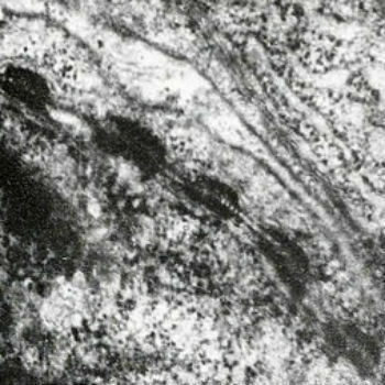
The black plates are the desmosomes observed under a microscope
They are shaped like a circular plate and merge into another identical structure on the surface of the nearest cell. We can compare the desmosomes to a push-button, made up of two complementary halves that fit together, one in each cell. Thus, when they join, they join adjacent cells.
Also read about the epithelial tissue.
How do desmosomes hold cells together?
A desmosome is characterized by two circular plates of proteins, one in each cell. From each plate, protein strands depart which cross the plasma membrane and occupy the intercellular space, where they associate with the protein strands of the adjacent plate.
The junction between adjacent cells is mediated by transmembrane proteins of the cadherin group. The long peptide chain of the cadherins sticks out of the cell and attaches to the ends of the cadherins in the adjacent cell.
The association of the filaments is what holds the two plates together, allowing the cells to be tightly connected.
In addition, the desmosome plates are made up of proteins (desmoplakins, placoglobins), which cross the membranes and stick the cells in the contact region.
Meanwhile, the part of the cadherin chain that faces into the cell binds to the intermediate strands rather than to the actin strands. The desmosomes are also attached to the filaments of another protein, keratin. This allows the anchoring of the desmosome to the cellular structure.
You hemidesmosomes, resemble desmosomes, but have different structure and function. They connect the plasma membrane of the epithelial cells to the adjacent basal lamina through keratin filaments. In hemidesmosomes there are no cadherins, but integrin proteins.