O heart is an organ of the Cardiovascular system responsible for pumping the blood and ensure that, in this way, it reaches the whole body. The human heart has four cavities: two atria and two ventricles. The number of cavities can be a criterion to differentiate the heart from the groups of vertebrates. Blood reaches the heart through the atria and leaves the heart through the ventricles.
Read more: Human body - main organs and systems
Human heart
The human heart is located between the lungs and behind our sternum, being therefore protected by our rib cage. the heart has the size of a clenched fist and weighs about 300 grams, and, despite being relatively small, it plays a big role in our body: the pumping of blood, thus providing that nutrients and oxygen reach the cells and wastes from metabolism can be taken to the proper place for disposal.

The heart has a cycle of contraction and relaxation. When it contracts, blood is pumped, and when it relaxes, it enters your cavities. The contraction phase is called
systole, and the relaxation one, diastole.The human heart is basically formed by muscle tissue cardiac striatum, a tissue of involuntary contraction. The heart wall is formed by three layers: the inner, also called the endocardium; the average, also called myocardium; and the external, also called epicardium. Striated cardiac muscle tissue is found forming the myocardium.
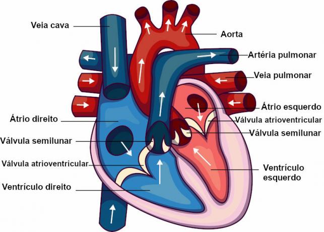
The human heart has four cavities: two atria and two ventricles. Blood enters the heart through the atria and leaves it through the ventricles. Upon reaching the heart, it flows to the ventricles, and from there it is driven to other parts of the body. The walls of the ventricles are thicker, and their contraction is much more vigorous than that of the atria, which is a way to guarantee the necessary impulse.
The heart also has valves, which prevent the backflow of blood. It features two valves atrioventricular, between the atrium and the ventricle, and two semi-lunar valves, at the exits of the heart.
blood path through the body
The blood, to complete a complete circuit through our body, passes twice through the heart (double circulation). The circuit that blood makes from the heart to the lungs with its return to the heart is called the small circulation or pulmonary circulation. The path that blood takes from the heart to the rest of the body with its return to the heart is called systemic circulation or large circulation.

O blood reaches the heart, through vein superior and inferior cava in the right atrium. This blood is oxygen-poor and comes from different parts of the body (excluding the lungs). The blood present in the right atrium goes to the right ventricle, which pumps it towards the lungs.
Blood travels to the lungs through the pulmonary arteries. Upon reaching them, oxygen-poor blood receives oxygen from breathing and becomes oxygenated. the oxygen rich blood return to heart through the pulmonary veins, being released into the left atrium.
The blood goes from the left atrium to the left ventricle. The left ventricle is responsible for pumping it to the body (excluding the lung). The blood goes through the aorta artery, what it branches to the capillaries. In capillaries, the oxygen present in the blood passes to the tissues and the carbon dioxide produced in the cellular respiration, from the tissues passes into the blood. The capillaries unite forming venules, which carry blood into the veins. The superior and inferior vena cava flow into the right atrium, restarting the cycle again.
Read more: Circulation of vertebrates - differences between groups
vertebrate heart
Different groups of vertebrates have differences in the anatomy of the heart. We can differentiate the heart of each group by analyzing the number of cavities it has. See, below, what the heart of each group is like:
- Fish: the heart of fish has only two cavities: an atrium and a ventricle. In the hearts of these animals, only oxygen-poor blood circulates.
- amphibians: the amphibian heart has three cavities: two atria and only one ventricle. In this heart, blood that is poor and rich in oxygen circulates, and the two types meet in the ventricle.
- reptiles: in the case of reptiles, we can observe differences within the group itself. While non-crocodilian reptiles have a heart with three cameras, the ventricle being partially divided, in the crocodilian reptiles, the presence of four cavities is observed: two atria and two ventricles. In the heart of reptiles, there is the passage of blood rich and poor in oxygen. In non-crocodilians, oxygen-rich blood meets oxygen-poor blood in the ventricle region. In crocodilians, on the other hand, the complete division into four cameras prevents blood from meeting in this location, but it occurs at the exit of the heart.
- birds and mammals: in birds and mammals, we observe a heart with two completely separate atria and two ventricles. In these animals, the left side of the heart receives and pumps only oxygen-rich blood, while the right side only passes oxygen-poor blood. If you want to know more about the topic of this topic, read: vertebrate heart.


