Ribosomes are structures, related to protein synthesis, that occur in all types. cell phones, even in prokaryotes. Free in the cytosol, or associated with membranes, ribosomes are essential for the individual's cell function and survival. See more about this important structure below.
What are ribosomes?
These are small particles measuring from 20 nm to 30 nm, without membranes and formed by proteins and ribosomal RNA (rRNA). Some authors claim that ribosomes are non-membranous organelles, others, however, assume that they, due to the absence of membranes, cannot be considered cell organelles.
US eukaryotes, the ribosomes are formed by four types ofribosomal RNA and approximately 80 proteins many different. Most ribosomal RNA is produced in the nucleolus, while proteins are produced in the cytoplasm. The proteins that will form the ribosome migrate from the cytoplasm to the nucleus and associate with ribosomal RNA, forming subunits, which migrate to the cytoplasm.

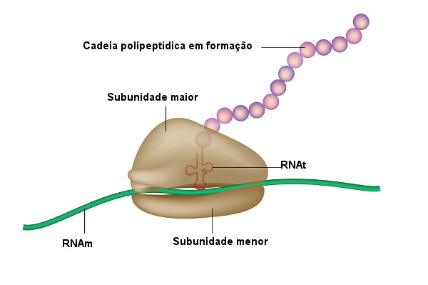
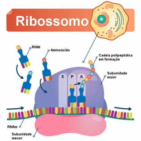
Ribosomes are formed by two subunits: major subunit and smaller subunit. These leave the nucleus separated, but unite in the cytoplasm. A functional ribosome is formed by both joined and linked to a messenger RNA molecule (mRNA).
This functional ribosome is responsible for ensure protein synthesis, and when the complex is formed, we can observe thatfour distinct binding sites, one site in the smaller unit and three sites in the larger unit (P, A and E sites):
Messenger RNA molecule binding site present in the smaller unit.
Site P: In this location, a transporter RNA molecule (tRNA) can be observed attached to the polypeptide chain that is being formed.
Site A: In this location, the presence of transporter RNA carrying the next amino acid, which will be attached to the polypeptide chain.
Site E: At this exit site, the unloaded transport RNAs leave the ribosome.
Read more: Types of RNA - fundamental molecule for protein synthesis
Do not stop now... There's more after the advertising ;)
Where are the ribosomes located?
Ribosomes can be found free in the cytosol (free ribosomes) or else membrane boundendoplasmic reticulum and nuclear envelope (linked ribosomes).
We cannot forget about the ribosomes found inside chloroplasts and mitochondria and that stand out for being smaller than the others mentioned. In prokaryotic cells, where there is no defined nucleus or membranous organelles, ribosomes are found only free in the cytosol.
function of ribosomes
Ribosomes are organelles responsible for protein synthesis in the cell. Cells responsible for large production of proteins, such as those of the pancreas, are rich in these structures. Furthermore, in cells with high metabolic activity, ribosomes can be found in clusters, known as polyribosomes.
Regardless of where the ribosomes are in the cell (free or bound), they will act in protein synthesis. The main difference, however, is in the destination of these proteins. Ribosomes present in the cytosol produce proteins that are usually destined for the cytosol itself. Linked ribosomes, on the other hand, usually synthesize proteins that will be inserted into membranes so that they can be packaged or secreted by the cell.
Also access:Proteins - macromolecules very important for living organisms
protein synthesis
Protein synthesis can be divided into three steps: start, elongation and end.
At start step, the approximation of messenger RNA and transporter RNA molecules is observed, in addition to the ribosome subunits. The transporter RNA, in this step, will take the first amino acid that will form the polypeptide chain.

After the start step, we have the elongation step. Here the amino acids are added one by one. The transporter RNA arrives at the A site and pairs by complementarity with the messenger RNA codon. A peptide bond then occurs between the amino acid at the A site and the forming polypeptide chain at the P site. The ribosome moves the transporter RNA from the A site to the P site, and the transporter RNA, from the P site, goes to the E site, where it is released. Messenger RNA also moves in this process, causing site A to locate at the next codon that will be translated.
The last step is the finishing step, marked by the arrival at site A of the termination codon ribosome. The UAG, UAA and UGA cracks signal the end of translation, as they do not encode any amino acid. When these cracks appear, the release factor, that will be responsible for releasing the polypeptide. After the end of the process, all the components separate, including the two ribosomal subunits. To learn more about the protein synthesis process, access the text: protein synthesis.
Difference between prokaryote and eukaryote ribosomes
the ribosomes of prokaryotes and eukaryotes they are very similar in structure, however, small differences can be observed between them. Generally speaking, the ribosomes of eukaryotes are larger than those present in prokaryotic organisms. In addition, there is a small difference with regard to composition, with the ribosomes of more complex eukaryotes.
By M. Vanessa Sardinha dos Santos
Biology teacher


