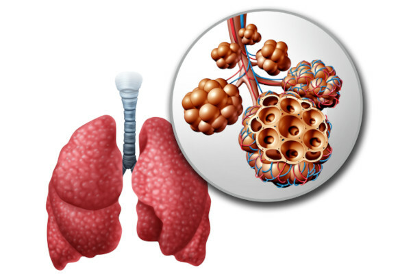
pulmonary alveoli they are structures that resemble small bags and have very thin walls. Wall thickness is directly related to the function of these structures, which is to carry out gas exchange. The pulmonary alveoli give the lung a spongy appearance.
Read more: Respiratory system - ensures oxygen capture and carbon dioxide release
What are pulmonary alveoli?
The pulmonary alveoli are small sacculiform structures which are arranged in a similar way to a beehive comb. These are the alveoli that form most of the lung parenchyma and provide a spongy aspect to the organ. They constitute the last part of the bronchial tree, being found in the alveolar sacs (structures formed by several alveoli), alveolar ducts and respiratory bronchioles.

Structure of the pulmonary alveoli
The alveoli are sacculiform structures that present thin walls formed by a layer of epithelial tissue that rests on a connective tissue, which features a large quantity of blood capillaries.
The alveolar wall is common to two adjacent alveoli, forming what we call the interalveolar septum. This septum is formed by two layers of cells called pneumocytes separated by connective tissue and blood capillaries. The interalveolar septum has pores that communicate with adjacent alveoli, this communication allows to balance the pressure difference.
Do not stop now... There's more after the advertising ;)
Pneumocytes can be type I or type II. O type I pneumocyte it is also known as a squamous alveolar cell and has a flattened nucleus. Its main function is to form a barrier that prevents the passage of extracellular fluid into the alveoli, but guarantees the occurrence of gas exchange. This passage is facilitated due to the thin cell thickness.
O type II pneumocyte it is also called septal cell, has a spherical nucleus and is located between type I pneumocytes. Type II pneumocytes are responsible for the secretion of pulmonary surfactant, which has the function of reducing the surface tension of the alveoli, reducing the force for inspiration and ensuring greater ease in breathing. Without the surfactant, which is a mixture of phospholipids, proteins and ions, the alveoli could collapse during exhalation.
Inside the interalveolar septa and on the surface of the alveoli, the alveolar macrophages, which ensure that invading microorganisms and other particles are phagocytosed. We can say that macrophages work by clearing the lungs of particles that were not eliminated in other parts of the airways.
Read too: What is Phagocytosis?
What is the function of the pulmonary alveoli?

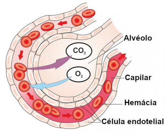
The pulmonary alveoli are a site of performing gas exchanges, in which carbon dioxide, present in the blood, passes to the interior of the alveoli, and oxygen, present in the inspired air, passes from the interior of the alveoli to the blood. This process is known ashematosis.
The oxygen that reaches the blood binds to the hemoglobin gives Red blood cell and is distributed to every cell in the body, where it will be used in the process of cellular respiration. On the other hand, carbon dioxide, which diffuses into the alveolus, is taken to the external environment by the airways in the expiration process.
Four structures must be traversed by the gases for gas exchanges to take place: cytoplasm pneumocyte, pneumocyte basal lamina, capillary basal lamina, and endothelial cell cytoplasm. Although it seems like a long way, the thickness of these structures is from 0.1 µm to 1.5 µm.
Read more: Systemic and pulmonary circulation
Pulmonary emphysema
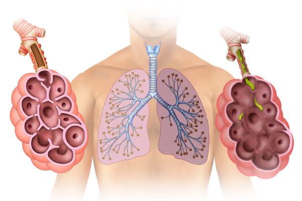
Pulmonary emphysema, according to the Primary Care Notebooks - Chronic Respiratory Diseases, of the Ministry of Health, is defined anatomically as an increase in the air spaces distal to the terminal bronchioles, with destruction of the alveolar walls. In this disease, therefore, the pulmonary alveoli are compromised and the consequent reduction in the body's capacity to carry out gas exchange.

The main cause of pulmonary emphysema is the use of cigarette, but other factors can lead to the development of the disease, such as atmospheric pollution. Symptoms include chronic cough, shortness of breath and recurrent respiratory infections.
The disease has no cure, but certain medications and rehabilitation therapy can improve the patient's quality of life. It is important that, in the case of smokers, the use of cigarettes is stopped, in order to prevent the disease from worsening. If you want to know more about this condition, read: Pulmonary emphysema.
By Vanessa Sardinha dos Santos
Biology teacher
Would you like to reference this text in a school or academic work? Look:
SANTOS, Vanessa Sardinha dos. "Lung alveoli"; Brazil School. Available in: https://brasilescola.uol.com.br/biologia/alveolos-pulmonares.htm. Accessed on June 27, 2021.


