You X ray they are electromagnetic radiation frequency, produced from the collision of beams of electrons with metals. This radiation cannot be perceived by the human eye as it is beyond the maximum frequency distinguished by human vision. It is important in Medicine because it makes it possible to generate diagnoses through images.
Discovery
In November 1895, the German physicist Wilhelm Conrad Roentgen (1845-1923) discovered X-rays while performing experiments in his laboratory. Using a cathode ray tube, Roentgen observed an unexpected luminosity and, interrupting it with his hand, saw the image of his bones exposed on a screen.
The physicist observed that the radiation, called X-rays, was capable of blackening photographic films. Then, in December 1895, he asked his wife, Anna Bertha Roentgen, to place her hand between photographic film and the tube from which the rays were produced. After a while, he noticed that the image of the woman's hand bones was imprinted on the photographic film. This was the first radiograph taken in the world. In 1901, Wilhelm Conrad Roentgen won the prize
Nobel Prize in Physics for his discovery.In Brazil, the first radiography was performed in 1896.
How are X-rays produced?
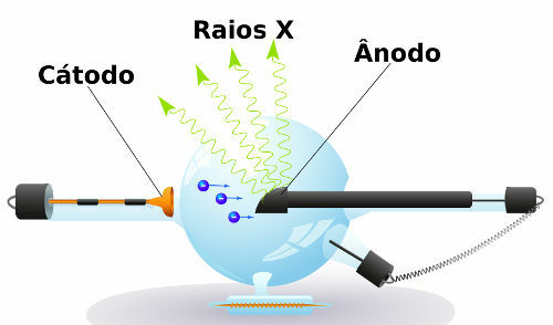
In a cathode ray tube, the cathode, after being heated by the passage of electric current, releases electrons at high speed. These electrons are strongly attracted by the anode, in which they end up colliding, as can be seen in the diagram below:

When the electrons of the atoms belonging to the anode receive energy from the moving electrons, the result is the production of electromagnetic radiation, which is called X-rays.
Do not stop now... There's more after the advertising ;)
X-rays, like all electromagnetic radiation, do not need a propagation medium and move in the speed of light (3.0 x 10⁸ m/s). That radiation is ionizing, therefore, it can cause damage to the human body in case of long-term exposure. The intensity of the X-rays is inversely proportional to the square of the distance, so the further away from the source, the lower the intensity of the rays.
X-ray exam
The great benefit arising from the discovery of X-rays was the possibility of performing diagnostic imaging. Tissues and muscle fibers they are practically crossed by x-rays, while the bones absorb this radiation. As these rays have the ability to blacken photographic plates, when placing parts of the human body between an x-ray source and a photographic plate, one can observe the formation of a "photograph" of the bones.

Radiography, or X-ray examination, is an example of how electromagnetic radiation can be used in medicine.
The study of organs of the abdomen, chest radiography for analysis of lung diseases and the mammography, exam that seeks to identify breast cancer, are examples of X-ray applications.
By Joab Silas
Graduated in Physics
Would you like to reference this text in a school or academic work? Look:
JUNIOR, Joab Silas da Silva. "What are X-rays?"; Brazil School. Available in: https://brasilescola.uol.com.br/o-que-e/fisica/o-que-sao-os-raios-x.htm. Accessed on June 27, 2021.
Chemistry

Electromagnetic waves, Biometrics, biological traces, fingerprints, hand venous structure, iris shape, security alarms, motion detectors, electric and magnetic fields.

